



服务热线
15365833739 
Dual-energy X-ray Absorptiometry (DXA) has long been recognized as the reference standard for bone mineral density measurement in clinical screening, diagnosis and follow-up management of osteoporosis. Nevertheless, conventional fixed DXA systems feature bulky gantries and scanning tables, restricting their installation to dedicated radiology rooms only. This creates difficulties in delivering timely testing for critically ill, post-operative bedridden or mobility-impaired patients. How can gold-standard precision break free from fixed examination rooms and deliver diagnostic-grade data within confined spaces such as ward bedsides? The portable DXA-500A Bone Densitometer offers a practical, sophisticated solution.
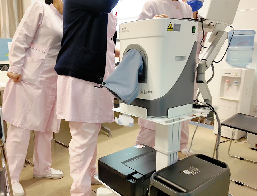
Portability Without Compromise: Reengineered Core Technology
The compact footprint of the DXA-500A Dual-energy X-ray Bone Densitometer does not come at the cost of diagnostic performance. The device retains the WHO-recommended dual-energy X-ray absorptiometry as its fundamental measuring principle. Building upon this foundation, the R&D team integrated cone-beam planar imaging technology, which captures the entire region of interest (ROI) in a single exposure. Paired with proprietary compensation algorithms that perform real-time correction for soft tissue, bone margins and scattered radiation signals, the total scanning time is controlled within 4 seconds. This design minimizes motion artifacts caused by involuntary limb movement in patients and enables high-throughput population screening.
Notably, engineers retained the complete X-ray generation and detection chain despite the highly compact chassis. By optimizing optical path layout and detector dynamic range, the DXA-500A captures raw data with sufficient signal-to-noise ratio under low radiation dose, guaranteeing precise and reproducible bone mineral density readings.
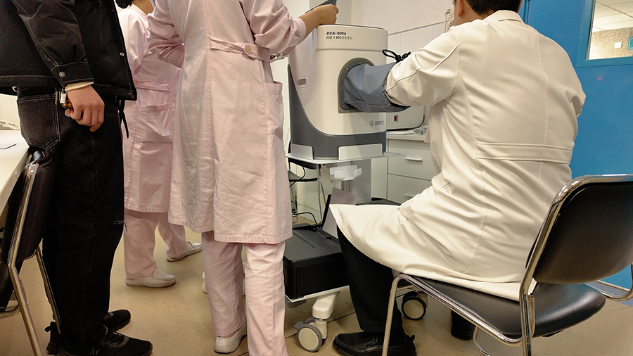
Triple Positioning Assurance: Consistent Stability On the Move
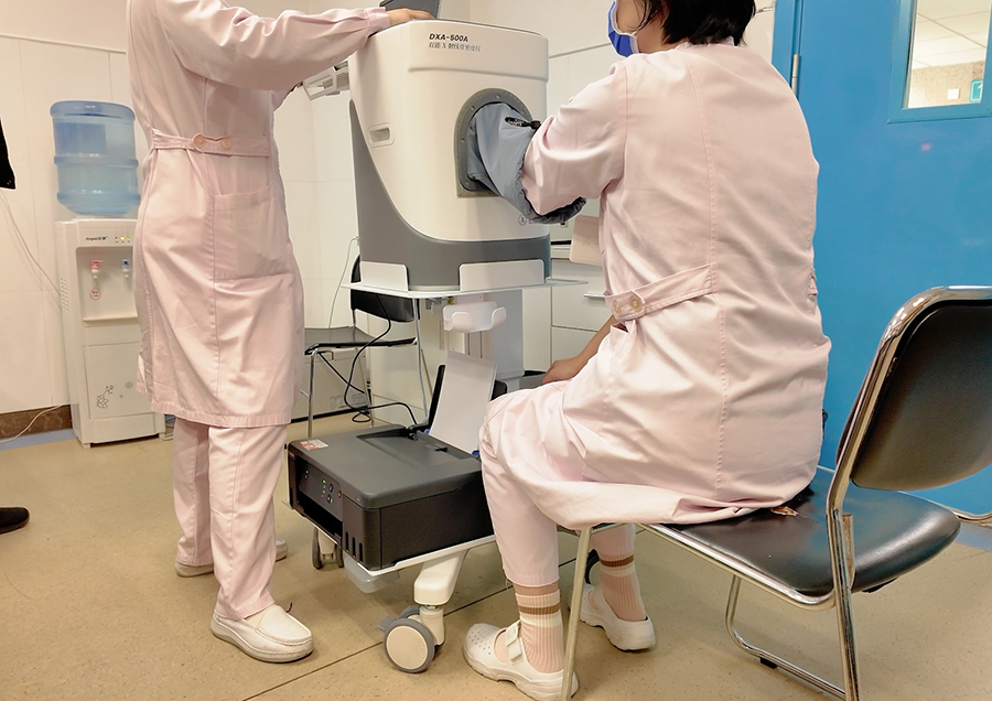
A key challenge facing portable systems is maintaining consistent patient positioning without a dedicated fixed scanning table. The DXA-500A incorporates a triple safeguard system: physical ergonomic grip, real-time video monitoring and laser alignment guides.
Physical Ergonomic Grip: The unit integrates forearm-biomechanical handles, stabilizing the patient’s forearm in natural seated or supine postures to reduce positional shifts from voluntary movement.
Real-time Video Monitoring: Live footage displays the patient’s limb posture, allowing operators to remotely assess critical parameters including elbow flexion angle and palm pronation, with instant corrective adjustments available.
Laser Alignment Guides: High-visibility laser lines enable rapid targeting of the distal radius and ulna, ideal for variable lighting conditions at patient bedsides.
Combined, these three mechanisms drastically reduce positioning deviation. For patients requiring long-term serial follow-ups, consistent positioning greatly improves comparability between sequential scans. Clinicians no longer face diagnostic interference stemming from posture variations across different operators or testing sessions.
Auto-Recognition Technology: Streamlined Workflow From Scan to Interpretation
The clinical value of bone densitometry extends far beyond absolute numerical values; rapid identification of skeletal abnormalities is equally critical. The DXA-500A features an automatic ROI recognition algorithm for the distal radius and ulna. Post-scan, three contouring modes are available for operator selection: single-point positioning, manual tracing and auto-tracing. Operators may select the optimal workflow based on image clarity and clinical preference to generate reliable bone mineral density results efficiently.
This architecture balances automated efficiency with manual adjustability: auto-tracing delivers instant ROI segmentation for standard high-quality scans, while single-point positioning and manual tracing deliver fine-grained control for complex cases with skeletal lesions or imaging artifacts. Radiology technologists are relieved from repetitive, labor-intensive manual edge outlining, streamlining daily clinical workflows.
Diagnostic-Grade Data Delivered Directly to Ward Bedside
The DXA-500A does not generate compromised, portable-only reference values; it produces diagnostic-grade data identical in reliability to fixed DXA systems, fully compliant with domestic and international quality control standards for bone densitometry. Validated via phantom calibration and multi-center clinical trials, its precision and accuracy metrics fully meet the specifications laid out in osteoporosis clinical practice guidelines.
This compact unit can be effortlessly wheeled into orthopedics, geriatrics, endocrinology wards and even mobile health screening vehicles, completing rapid forearm scans without repositioning bedridden patients. Its performance is enabled by deep integration of cone-beam imaging, signal compensation algorithms and ergonomic positioning engineering. For department directors and radiology technologists, the DXA-500A is more than a space-saving upgrade — it delivers a viable pathway to preserve the gold-standard DXA measurement accuracy at patient bedsides. Freed from the confines of dedicated scanning rooms, precise bone density testing can now reach a far broader spectrum of clinical scenarios.